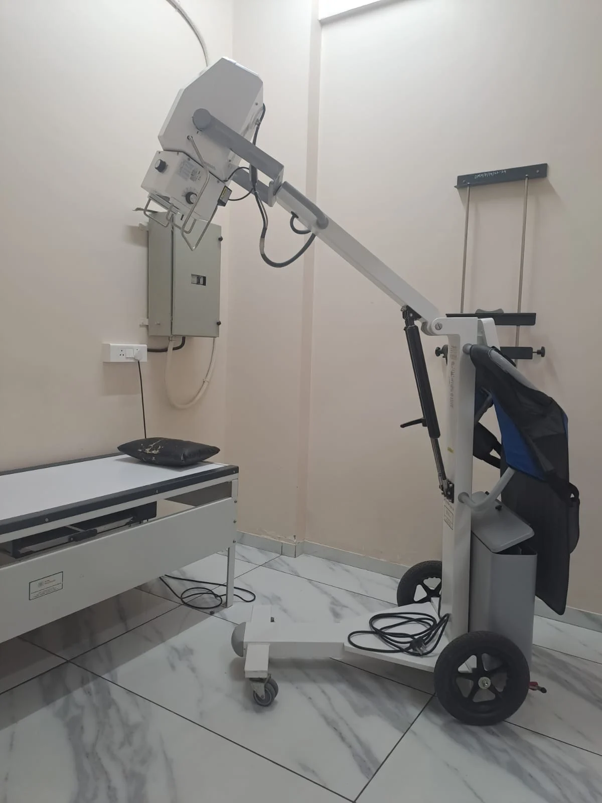
What a digital X-ray can show
An X-ray creates images using a small amount of ionizing radiation and can help assess selected bones or other structures. It is useful for some questions but does not show every tissue or explain every symptom. The clinician chooses the body area and views based on the concern being investigated.
Ask what the X-ray is intended to clarify and whether the result may change treatment. A normal image does not always rule out every problem, and an abnormal finding may need context. The clinician interprets the report alongside your symptoms and examination.
Preparing for the examination
Most X-rays require little preparation, but instructions can vary by body area and department. Staff may ask you to remove jewellery or clothing that could obscure the image. Tell them if you need help standing, sitting, moving or holding a position so they can plan safely.
Tell the radiographer if you are pregnant or may be pregnant before the examination. Bring prior reports or mention earlier imaging of the same area. Do not assume that a previous image is available to a new team unless you confirm how it can be accessed.
Positioning, comfort and image quality
The radiographer may take more than one view and ask you to stay still briefly. Positioning helps produce an image that can answer the clinical question. If a position causes significant pain or is not possible, tell staff rather than forcing the movement.
The team may repeat an image if movement or positioning makes it difficult to interpret. This is a technical decision made by staff. Ask for an explanation if you are concerned about the number of views or the process.
Radiation safety and personal circumstances
Because X-rays use ionizing radiation, the team considers whether the examination is justified for the clinical question. Tell staff about pregnancy or possible pregnancy, and mention any recent imaging if relevant. Do not delay a necessary examination without discussing the risks and benefits with the referring clinician.
Protective measures and technique are selected by qualified staff according to the examination. Ask the radiographer if you have a safety question. Advice can vary based on body area, age and clinical need.
Receiving and discussing results
A radiologist or qualified clinician may review the images and issue a report. Ask when it will be available and who will explain it. Do not assume that a technician’s brief comment is the final diagnosis or that no follow-up is needed if the image was taken quickly.
Keep the report with your records and share it with the clinician managing the concern. If the report recommends another step, ask what it means for you and how soon to act. Seek earlier assessment if symptoms worsen before the planned discussion.
Questions to ask at the imaging visit
Before leaving, confirm that the requested body area was imaged and ask how results are returned. Tell staff if you need an accessible explanation, mobility assistance or an interpreter. If you are uncertain about a preparation or safety instruction, ask before the examination begins.
The X-ray page is general information and cannot determine whether imaging is appropriate for your symptoms. That decision belongs to a clinician who can review your situation. For sudden severe symptoms or serious injury, seek urgent care rather than waiting for a routine imaging slot.
Imaging children and people who need assistance
Tell staff if the patient is a child, has difficulty standing or needs a caregiver nearby. The radiographer can explain positioning and discuss practical support. Pregnancy or possible pregnancy should be raised before an X-ray so the team can consider the appropriate approach.
A support person may need to follow protective instructions or wait outside the room. Ask the department what is permitted and whether a different arrangement can be made. Do not hold a child or equipment unless staff instruct you to.
Understanding a request for more images
A radiographer may take additional views if the first images do not answer the clinical question. If this happens, ask for a brief explanation. The clinician reviewing the result decides what it means and whether further assessment is needed.
If another X-ray is requested later, mention the date and location of the earlier study. Sharing previous images may help comparison. Tell the clinician if you are concerned about repeat exposure so the reason can be discussed.
What happens if the X-ray does not explain pain
Some problems are not visible on an X-ray or may need a different test. If symptoms continue, the clinician may review the examination, image and timeline and decide whether additional assessment is useful. A normal X-ray should not be treated as proof that symptoms are imaginary.
Ask what to do if pain, swelling or function changes and when follow-up is needed. If there is severe injury, loss of sensation or circulation, or sudden inability to use the area, seek urgent assessment.
Using X-rays as one part of an assessment
A digital X-ray can help answer selected questions, but it is not a complete examination of every structure. If pain continues despite a normal result, the clinician may reassess symptoms or consider another test. Ask what the image has ruled in or out and what remains uncertain.
Do not conclude that an injury is minor solely because an initial image was normal. Follow the advice given and arrange review if pain, swelling or function worsens. Sudden severe symptoms or loss of sensation require prompt assessment.
Sharing images with another clinician
If you are seeing another clinician, ask how to share the report and original images. A report alone may not answer a later question, and image access can help comparison. Keep the date, body area and facility details with the files.
Use a secure method approved by the hospital for transferring medical records. Do not post X-rays or reports publicly. If the image cannot be accessed, ask the imaging department what options are available.