






When a doctor recommends a brain scan, it is natural to wonder why one test has been chosen over another. CT and MRI both produce images of the brain, but they work differently and answer different clinical questions. Neither is universally “better” in every situation. The right choice depends on the symptoms, how urgently an answer is needed, and which structures or abnormalities the medical team needs to examine. This article explains CT vs MRI for brain tumour evaluation in straightforward terms, including how each test works, what to expect, and what happens if a scan shows an abnormality.
What is a CT scan?
Computed tomography (CT) uses X-rays taken from different angles and a computer to create cross-sectional images. The scan is quick and can show the brain, skull, swelling, many types of bleeding and some tumours. Because it can be completed rapidly, CT is often used first in emergency situations such as suspected bleeding, significant head injury or possible stroke. CT does involve ionising radiation. For a medically necessary scan, clinicians weigh the expected diagnostic benefit against the small potential risk associated with radiation exposure. Tell the doctor and imaging team if you are pregnant or could be pregnant.
What is an MRI scan?
Magnetic resonance imaging (MRI) uses a strong magnetic field, radio waves and a computer to produce detailed images. It does not use ionising radiation. MRI is especially useful for evaluating brain tissue and is commonly used when a suspected tumour needs a closer look at its size, position and relationship to nearby structures. Some MRI examinations use a contrast agent, often gadolinium-based, to help distinguish tissues and characterise abnormalities. Contrast is not required for every scan. The clinician decides whether it is useful and checks relevant safety factors.
CT vs MRI for brain tumour: key differences
| Feature | CT scan | MRI scan |
|---|---|---|
| How it works | X-rays and computer processing | Magnetic field and radio waves |
| Speed | Usually faster | Usually takes longer |
| Emergency use | Often useful when a rapid answer is needed | Useful in selected urgent and planned assessments |
| Soft-tissue detail | Good for many urgent findings | Usually provides more detailed brain-tissue images |
| Bleeding and skull injury | Particularly useful in many emergency settings | Can add detail in selected cases |
| Suspected tumour | Can identify many abnormalities | Often preferred for detailed tumour assessment |
| Radiation | Uses ionising radiation | Does not use ionising radiation |
| Main practical limits | Radiation exposure; some findings need further imaging | Longer scan, noise, enclosed space and implant-safety checks |
This comparison is a guide, not a rule for every patient. The medical team may use one test first and another later because they provide complementary information.
When might a CT scan be preferred?
A possible brain bleed or acute emergency
If a person develops a sudden severe headache, new neurological weakness, a seizure or reduced consciousness, clinicians may need to identify bleeding quickly. CT is often the first investigation because it is fast and widely available in emergency pathways. A suspected stroke also requires urgent assessment; do not wait to arrange a private scan if symptoms are sudden.
Head injury
CT can help detect skull fractures and acute bleeding after significant head trauma. Whether imaging is needed depends on the injury, symptoms, examination and clinical risk factors.
When MRI is not immediately suitable
Some implanted devices or metallic foreign bodies may make MRI unsafe or require specialist checks. In these situations, CT may provide important information. Modern MRI safety decisions are device-specific, so patients should share exact implant details rather than assuming every implant rules out MRI.
When might MRI be preferred?
A suspected brain tumour
MRI generally gives more detailed views of brain tissue. It can help show a lesion’s location, size, surrounding swelling and relationship to important structures. Specific MRI sequences may help the specialist narrow the possibilities, although imaging alone does not always establish the exact tumour type.
Persistent neurological symptoms
When symptoms and examination suggest a problem involving brain tissue, a clinician may recommend MRI to investigate further. The decision depends on the symptom pattern and possible diagnoses; not everyone with a headache needs an MRI.
Planning treatment or follow-up
If an abnormality has been identified, MRI may be used to clarify its features, support treatment planning or monitor changes over time. The follow-up schedule depends on the diagnosis and the specialist’s recommendation.
Does MRI always detect more than CT?
MRI usually provides better soft-tissue detail, but it is not the best first test for every problem. A fast CT may be more useful when a time-critical bleed must be identified. Some conditions are visible on both tests, while others require particular imaging sequences, contrast or additional investigations. A scan is only one part of diagnosis: symptoms, physical examination and medical history matter too.
Does a brain scan confirm that a tumour is cancerous?
No. CT or MRI may reveal a mass or another abnormality, but imaging does not always determine whether a tumour is benign or malignant. Some non-tumour conditions can also look like a mass. A radiologist interprets the images, and a specialist considers them alongside the clinical picture. In selected cases, a biopsy or surgical tissue sample is needed to establish the diagnosis. An abnormal result should be discussed with the doctor who requested the scan. Avoid interpreting a report in isolation or assuming the worst from unfamiliar terms. The doctor can explain which findings are important, whether additional tests are required and how quickly follow-up should occur.
What happens during a CT scan?
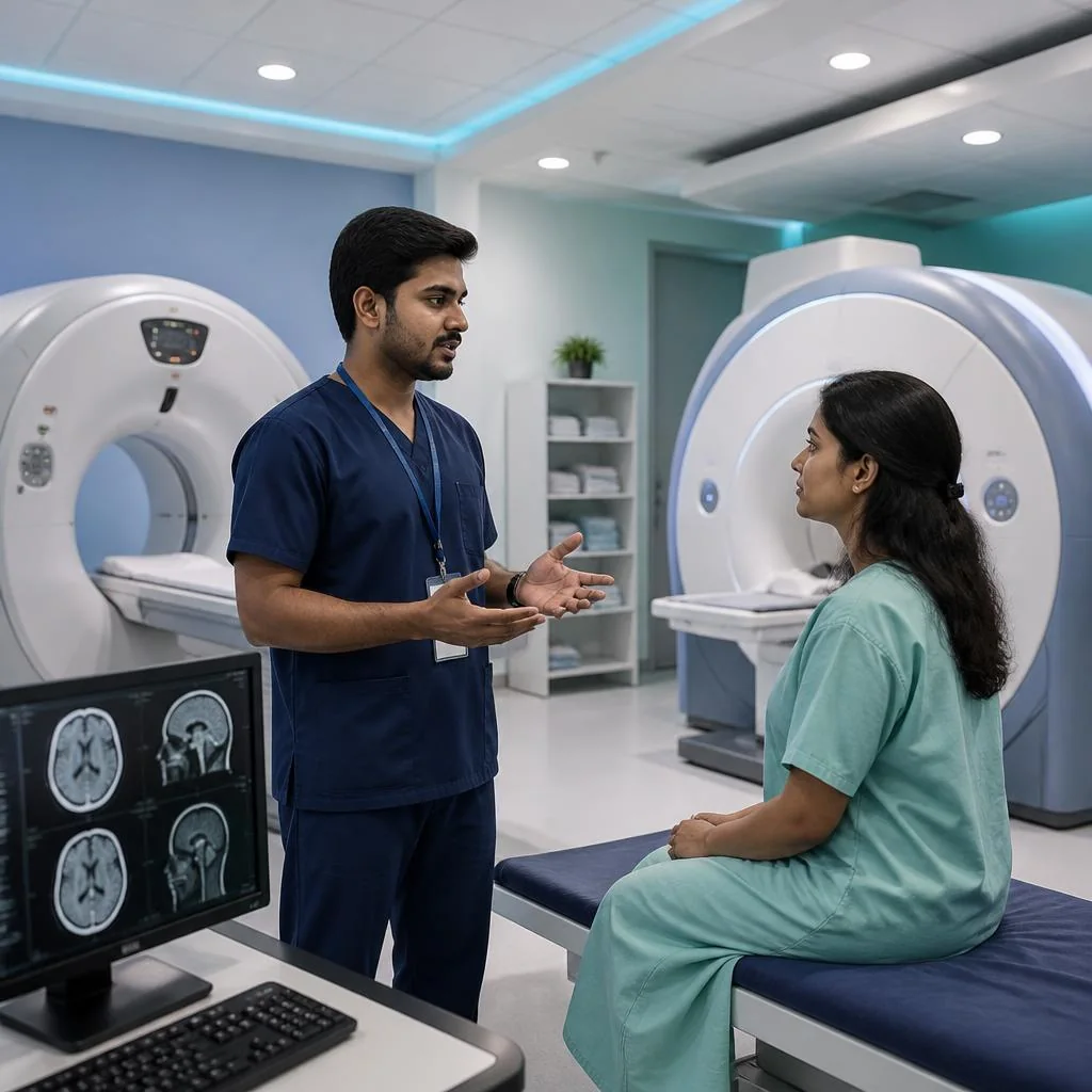
You may be asked to remove metal objects and lie on a table that moves through a ring-shaped scanner. The scan itself is usually brief, and the technologist may ask you to remain still. If contrast is planned, you will receive instructions about preparation and any relevant allergy or kidney-history checks. Follow the facility’s directions rather than changing medicines or meals on your own.
What happens during an MRI?
You lie on a table that moves into the scanner. The machine makes loud knocking sounds, so ear protection is provided. Remaining still helps produce clear images. Tell the team beforehand about pacemakers or other implants, metal fragments, previous surgeries, possible pregnancy, kidney disease if contrast is being considered, and anxiety in enclosed spaces. The staff will assess safety and discuss options if you are claustrophobic. MRI can feel long or uncomfortable for some people, but the imaging team can explain the process and help you prepare. Do not enter the MRI room with unapproved metal objects.
Risks and safety: what should patients know?
Both tests are widely used when clinically indicated. CT uses radiation, so repeated imaging should be based on medical need. MRI avoids ionising radiation but has important magnetic-field safety requirements. Contrast agents have their own considerations, including uncommon reactions and the need for additional assessment in some patients. Tell the medical team about allergies, kidney problems, pregnancy possibility and all implants or devices. The safest scan is not necessarily the one with the fewest technical risks; it is the appropriate investigation for the medical question, performed with suitable precautions.
How to prepare for your appointment
Bring prior reports and images, your current medication list, and a concise description of symptoms. Note when symptoms began, whether they are changing, and whether there have been seizures, weakness, speech changes, vomiting or vision problems. Ask why the scan is being recommended, whether contrast is planned, how results will be shared and who will explain them. If symptoms are sudden or severe, seek emergency care rather than waiting for a routine appointment or scan.
Brain imaging and specialist care in Sangli
For people in Sangli and nearby communities, Deccan Malti Neuro & Superspeciality Hospital in Vishrambag lists neurology, neurosurgery and diagnostic services including CT scan. A clinician can assess the concern and advise which investigation is appropriate. Confirm current test availability, preparation requirements and appointment details directly with the hospital. MRI availability should be checked before planning a visit.
Frequently asked questions
Which is better for a brain tumour, CT or MRI?
Is a CT scan faster than an MRI?
Generally, yes. CT is usually completed more quickly, which is one reason it is frequently used when urgent decisions are needed.
Does MRI use radiation?
No. MRI uses a magnetic field and radio waves, not ionising radiation. It still requires careful screening for implants and metal.
Can CT miss a brain tumour?
Some abnormalities are more clearly seen on MRI. If symptoms or the CT findings require further assessment, the doctor may recommend MRI or another test.
Is contrast always needed for a brain MRI?
No. Contrast depends on the clinical question and the information required. The clinician will decide whether it is appropriate and review relevant safety factors.
How soon will I receive the result?
Timing varies by facility and urgency. Ask the imaging centre when the report will be ready and arrange a follow-up with the clinician who requested the scan.
Where can I ask about brain imaging in Sangli?
Deccan Malti Neuro & Superspeciality Hospital is located in Vishrambag on Sangli–Miraj Road. Contact the hospital to confirm the current availability of the required investigation and specialist appointments.
Final takeaway
CT and MRI are complementary tools, not competing tests. CT is often valuable when speed matters; MRI usually provides greater detail for planned assessment of brain tissue and suspected tumours. The right decision comes from clinical assessment, not from choosing a scan independently.
Why the scan request may mention contrast
Contrast material can make some structures and abnormalities easier to distinguish. Whether it is needed depends on the suspected condition and the protocol chosen by the radiology team. If contrast is planned, share information about previous reactions to contrast agents, kidney disease, pregnancy possibility and relevant medicines. Do not assume contrast is dangerous or necessary in every case; ask what extra information it is expected to provide and follow the facility’s preparation instructions.
Understanding the radiology report
A radiology report describes what the images show and may include terms such as lesion, mass, swelling, enhancement or incidental finding. These words do not all mean cancer. The report should be interpreted by the clinician who knows why the scan was requested and what symptoms the patient has. Ask the clinician to explain the main finding, what remains uncertain, whether further imaging or a specialist review is recommended, and how quickly the next step should happen.
Why old scans are useful
Previous scans can help doctors identify whether a finding is new or has changed. Bring the actual images when possible, not only a written report, and tell the team where and when earlier tests were performed. Comparing images may avoid unnecessary repetition and can make follow-up more informative. Never delay emergency care to locate old records; urgent symptoms take priority.
Who interprets the images?
The radiologist is a doctor trained to interpret medical images and prepare a report for the clinician who requested the examination. The treating doctor then connects the findings with the patient’s symptoms, examination and medical history. Sometimes the report recommends another scan or specialist review because the first examination cannot answer every question. That does not automatically mean that something dangerous has been found; it means the next step may provide additional information. Patients should ask who will explain the result and how to contact that clinician. Do not make treatment decisions based only on an isolated phrase in a report or an online search. If the scan was performed because of sudden or worsening symptoms, follow the urgent instructions given by the treating team rather than waiting for a routine results appointment.
Ask about the next step
Before leaving, confirm whether the result will be reviewed by the doctor who requested the scan and whether a follow-up appointment is needed. If the report recommends additional imaging, ask what question the next test is intended to answer and how soon it should be arranged. Clear instructions help prevent delays and unnecessary worry.